Our Services
- Sound Therapy
- Psychology Internship
- Corporate Training
- Therapeutic Yoga and Meditation
- Emotional Freedom Technique
- Play Therapy
- Psychological Drama Therapy
- Creative Dance Therapy
- Art Therapy
- Medicinal Treatment
- Career Counseling
- NLP Training in India
- Hypnotherapy
- Psychotherapy in India
- Personality Development in India
- Yoga & Meditation Training
- Mind Power Training
- Psychological Counseling
- Psychometric Tests
- Remedial Classes for Learning Disability
- Brain Diagnosis
- EEG
- Alcoholism Treatment
Brain Diagnosis
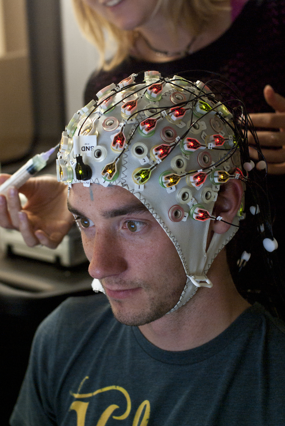
EEG (Electroencephalography) and brain mapping is a technique used to measure brain waves. It is a useful diagnostic tool for epilepsy, Encephalopathy, coma, SSPE, migraine variants, syncope, delirium, catatonia, sleep studies, various psychiatric illnesses, etc.
Clinical EEG is the depiction of electrical potentials from the Cerebrum. It is a non-invasive procedure. EEG has a high temporal resolution, on the order of milliseconds rather than seconds. EEG is commonly recorded at the sampling rates between 250 and 2000 Hz in clinical settings. It does not cause exposure to X-ray beams or too strong magnetic fields, hence safe in pregnant patients and those with pacemakers.
EEG is done by a trained technician. It involves pasting of electrodes on the scalp with the help of Kaolin powder paste. It does not cause any pain or electrical stimulation to the scalp. For this procedure, the patient should wash his/her head before coming to the clinic and keep it oil free. After the procedure, the paste can be easily removed from the scalp by washing the head. It is a good idea to bring a small napkin to wipe your head.
For very small children advised EEG by your doctor, they will be given a sedative medication to put them to sleep before the procedure. Normally they wake up within an hour after the procedure. It is wise to wake them early on the day of the procedure and do not allow them to sleep on the way to the clinic.child-patient.jpg In this way, they are already drowsy when they reach our place and we need to give them a minimum dose of the sleeping medication. Ensure that the head is kept oil-free and any cap or headgear is removed before the child sleeps. A normal EEG report does not exclude the possibility of a disease or disorder. It has to be taken in context with the clinical picture and other available data.
Dr. Manish Patel has undergone additional training in EEG reporting from National Institute of Mental Health and Neurosciences (NIMHANS), Bangalore.